Food's Journey Through Your Body
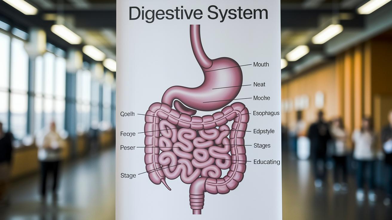
Ever wonder what happens to your food after you eat? This diagram shows you, step by step, how your food moves through your body.
Quick look:
• Food enters your mouth where it’s chewed.
• It travels through a system about 30 feet long.
• Each organ is clearly labeled to show its role.
The picture explains how chewed food turns into nutrients that keep you fueled every day. It's a simple, clear guide that breaks down the process in easy-to-follow steps.
Digestive System Diagram Labeled: Inspiring Clarity
This diagram gives you a clear, high-resolution look at your digestive system. It shows the full journey of food as it moves through your body so you can easily study how each organ works.
Your food starts in the mouth, where chewing breaks it down and saliva with amylase gets to work. Next, the esophagus uses a wave-like motion (peristalsis) to push the food toward the stomach.
In the stomach, which can hold about 1 liter, food mixes with acids and enzymes that help break it down further. The journey then moves to the small intestine, a long, 20‑foot tube, that absorbs most of the nutrients. After that, the large intestine reclaims water and packs up waste before it reaches the rectum for elimination.
This diagram also points out the role of accessory organs like the liver, which makes bile, and the pancreas, which releases enzymes to assist digestion. Together, these parts form an impressive system that can stretch to about 30 feet when laid out, with the biliary system playing a key role in the process.
Clear labels and short notes next to each element make it very easy to see what each part does. For a broader view, visit how does the digestive system work for more details on how your system keeps you going.
Major Organ Breakdown in the Labeled Digestive System Diagram
Your digestive system works like a well-tuned machine, breaking food into parts your body can use. Here’s what happens at each stage:
The Mouth
When you chew, your teeth shred food into smaller pieces. Saliva mixes in and contains an enzyme called salivary amylase that starts breaking down starch. Think of it like a food processor smoothing ingredients before cooking.
The Esophagus
A wave of muscle contractions, called peristalsis, pushes food down toward your stomach. It moves food like a gentle wave rolling in and carrying things along.
The Stomach
Here, food meets digestive acids and enzymes such as pepsin, which begin to break down proteins. The stomach churns your food into a semi-liquid mix and creates the best environment for these enzymes to work, much like a blender that heats and mixes at the same time.
The Small Intestine
This long tube is divided into the duodenum, jejunum, and ileum. In the duodenum, bile and pancreatic enzymes further break food into tiny molecules. The inner lining, rich in enzymes, quickly absorbs nutrients into the blood. Imagine a high-efficiency filter that pulls out every useful bit.
The Large Intestine
Undigested material moves into the large intestine, where water is reabsorbed to firm up stool. Friendly gut bacteria also help by fermenting some of the leftovers to produce short-chain fatty acids, much like a sponge soaking up extra water.
The Rectum
Waste is stored briefly in the rectum before it’s eliminated. Each organ has a specific role to ensure your body uses food efficiently.
For more details on how each part contributes, visit digestive system function.
Stepwise Labeled Food Transit in the Digestive System Diagram
Food enters your mouth, where chewing breaks it into a soft ball called a bolus, like kneading dough into a small ball.
Next, the bolus moves down the esophagus. Wave-like muscle actions, known as peristalsis (rhythmic squeezes), push it toward the stomach. In the stomach, acids and enzymes blend with the bolus to create a thick liquid called chyme, much like ingredients mixed in a blender.
When chyme reaches the duodenum, bile from the gallbladder and enzymes from the pancreas work to neutralize the acid. This sets up the food for further breakdown.
In the jejunum and ileum, your body absorbs important nutrients like proteins, fats, and carbohydrates, similar to how a sponge soaks up water.
Finally, in the large intestine, water is reclaimed and fibers break down through fermentation, and the remaining waste ends up in the rectum for elimination.
- Mouth: Chewing forms a bolus.
- Esophagus: Peristalsis moves the bolus, and the stomach turns it into chyme.
- Duodenum: Bile and enzymes neutralize stomach acid.
- Jejunum and Ileum: Nutrients are absorbed.
- Colon: Water is absorbed and fibers ferment.
- Rectum: Waste is prepped for elimination.
Clinician-reviewed • Last reviewed: 10/2023
Accessory Organs in the Labeled Digestive System Diagram: Liver, Pancreas, Gallbladder

Your digestive system chart shows three support organs that help with digestion.
The liver does many jobs. It makes bile to break down fat. It cleans your blood from toxins. It stores extra energy as glycogen. Imagine the liver as a processing plant that turns raw nutrients into useful supplies.
The gallbladder acts like a storage tank. It holds bile from the liver and sends it to your small intestine (duodenum) when you eat, especially fatty foods. This burst of bile breaks fat into smaller droplets that enzymes can digest easily.
The pancreas is an important helper. It makes enzymes like lipase (for fat), amylase (for carbohydrates), and proteases (for proteins) to break down food so your body can absorb it. It also sends bicarbonate to the duodenum to neutralize stomach acid. This protects your gut and helps enzymes work at the right pH.
- Liver: Bile making, blood cleaning, and energy storage.
- Gallbladder: Bile storage and release.
- Pancreas: Enzyme and bicarbonate production.
Together, these organs help you digest food better by breaking down fats and managing acidity.
Teaching and Learning with a Labeled Digestive System Diagram
This diagram is a simple tool that makes learning about the digestive system fun. Clear, color-coded labels help you see each organ and understand what it does. Teachers can use a child-friendly drawing to break down tough details and keep learners interested.
Fill-in-the-blank worksheets let students practice by labeling parts of the diagram. This active exercise helps lock in the information. Digital, interactive diagrams work well for online learning, letting you click on parts to see short notes on how each organ works.
Try these classroom strategies:
- Use color-coding to connect each organ with its function.
- Have students complete labeling worksheets to boost memory.
- Encourage drawing the diagram from memory to see how much they remember.
These techniques are useful for both quick reviews and deeper study sessions, making them a reliable resource for exam preparation and everyday learning.
Final Words
In the action, this post walked through each part of the digestive system using a clear, detailed digestive system diagram labeled. It explained how food moves from the mouth to the intestines, highlighted key roles of accessory organs, and offered ways to use the diagram to boost learning. The breakdown of each organ and its functions makes understanding digestion straightforward. Keep exploring and learning, every step helps you understand your body better.