
Quick Action
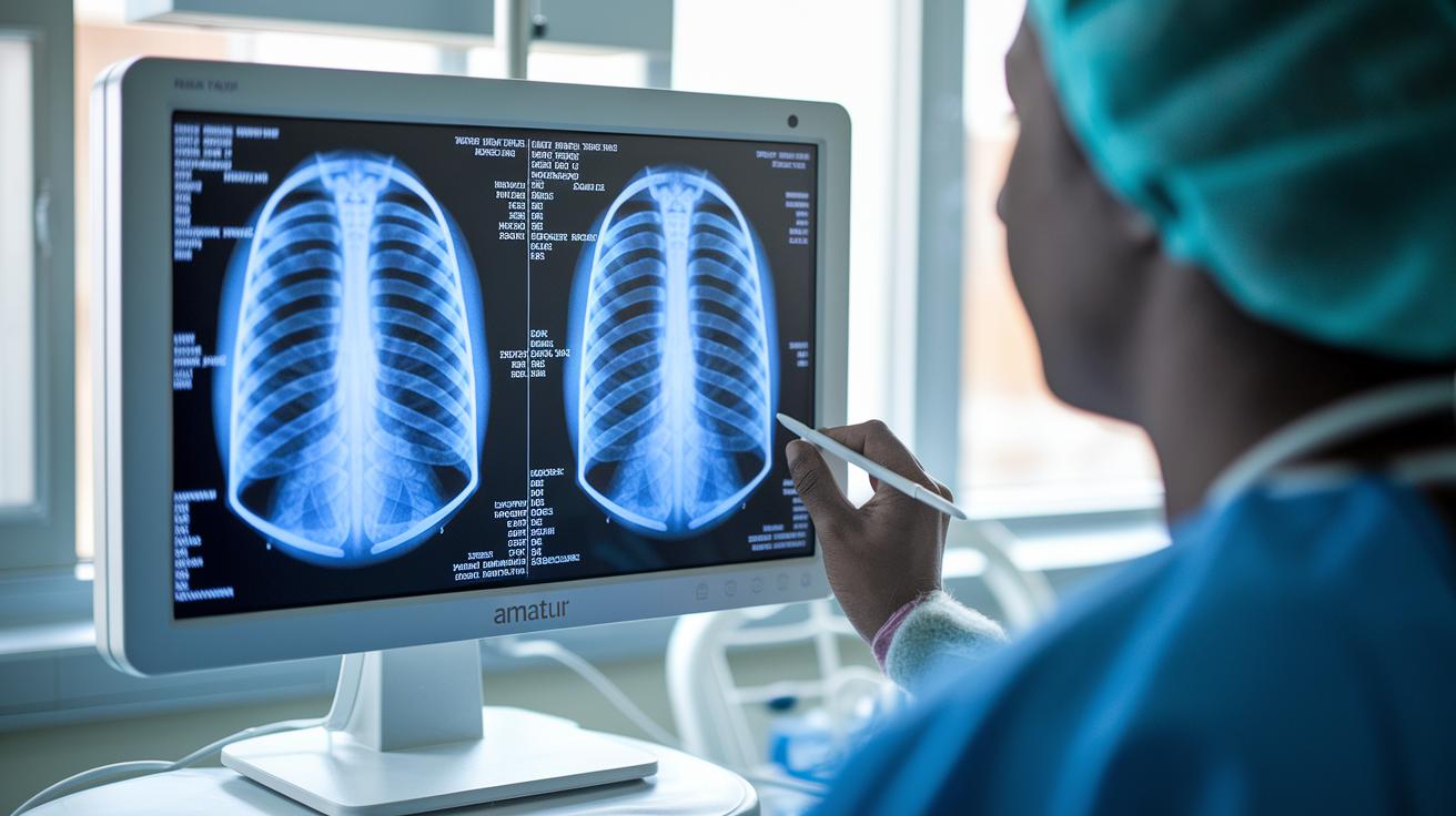
A chest x-ray can reveal hidden issues like sarcoidosis, even if you feel fine.
Red Flags
• Enlarged lymph nodes seen on the scan
• Tiny nodules in the lungs
What It Looks Like
Often, sarcoidosis shows up without any clear symptoms. The condition might only be noted when a scan unexpectedly finds changes in your chest.
Likely Causes
• The condition is usually discovered by chance during routine imaging tests.
• An underlying inflammation causes the lymph nodes to become larger or small nodules to form in the lungs.
What to Do Now
- Follow up with your doctor if your scan shows any unusual findings.
- Expect your doctor to review your health history and then order more tests, such as additional scans or a tissue biopsy (a small sample of tissue taken for study).
- Ask for a clear explanation about the tests and what the results mean for your treatment.
When to See a Clinician
If you have an abnormal chest x-ray, contact your healthcare provider as soon as possible to start further evaluation.
Clinician Reviewed: Yes | Last Reviewed: October 2023
Diagnostic Workflow for Confirming Sarcoidosis
Quick summary: Sarcoidosis affects about 200,000 US adults, mostly between the ages of 20 and 30. It is often discovered by chance during chest x-rays done for other reasons.
What it looks like: Many people with sarcoidosis have no symptoms. Routine imaging like x-rays can show signs such as enlarged lymph nodes or small lung nodules. Even if you feel fine, these findings call for further checks.
Step 1 – Get Your History and Do a Physical Exam:
Your doctor will ask when you first noticed any changes, what makes them better or worse, and look into your past health history. This conversation is the first clue in spotting sarcoidosis.
Step 2 – Do More Specific Tests:
After the initial check, more tests follow:
- Targeted imaging scans
- Laboratory tests
- Tissue biopsies (a small sample of tissue is taken to look for granulomas, which are tiny clusters of immune cells)
Each test helps the doctor rule out other conditions and put the pieces together for a clear diagnosis.
This step-by-step process is designed to remove uncertainty and confirm the diagnosis so that you can get the treatment you need.
Clinician-reviewed. Last reviewed: October 2023.
Role of Imaging in Diagnosis of Sarcoidosis
Quick action: Imaging tests help your doctor look inside your chest when sarcoidosis is suspected. They start with a simple chest X-ray and may move to more detailed scans if needed.
Red flags:
- Unclear findings on an X-ray.
- Signs of active inflammation or unusual patterns.
What it feels like: You may feel uneasy not knowing what is wrong. These tests give your doctor a closer look to guide the next steps.
Likely reasons for these tests:
- To check for enlarged lymph nodes.
- To spot lung nodules or small spots.
- To see patterns that might show scarring or unusual inflammation.
What to do now:
- Begin with a chest X-ray. It often shows enlarged lymph nodes and lung nodules.
- If the X-ray is not clear, ask about an HRCT scan. This test shows details like small nodules and fibrotic (scarring) changes.
- If your doctor is still unsure or if the disease may be active outside the lungs, a PET scan might be used. It helps show where inflammation is high and where a biopsy might be best taken.
When to see a clinician: If test results are unclear or you feel unwell, follow up with your doctor this week.
The doctor may ask when you first noticed symptoms and any changes in your breathing. They may also recommend a biopsy for a closer look.
| Modality | Key Findings | Use Case |
|---|---|---|
| Chest X-ray | Enlarged lymph nodes, lung nodules | Initial test and routine check |
| HRCT | Small nodules, fibrotic changes, unusual patterns | Detailed view when the X-ray is inconclusive |
| PET scan | Active inflammation, spots outside the lungs | Helps target biopsy areas and assess disease activity |
Clinician-reviewed (last-reviewed: 2023-10).
Laboratory Tests and Biomarkers for Sarcoidosis Confirmation
When your history, exam, and scans suggest sarcoidosis, your doctor usually orders blood tests next. These tests help build a fuller picture and track how active the illness is, though no single test on its own confirms the condition.
Key tests include:
- ACE level (elevated in about 60% of cases)
- Serum calcium
- Lysozyme
- Liver function tests
- Immunologic markers
An increased ACE level is a useful clue but can be normal in mild or inactive cases. Other markers like serum calcium, lysozyme, liver enzymes, and immune tests add more support for the diagnosis.
Remember, lab results are just one part of the puzzle. Your doctor will combine these results with imaging and sometimes a biopsy to make a final diagnosis.
Clinician-reviewed | Last-reviewed: October 2023
Biopsy and Histopathology in Confirming Sarcoidosis
Bronchoscopic Transbronchial Biopsy
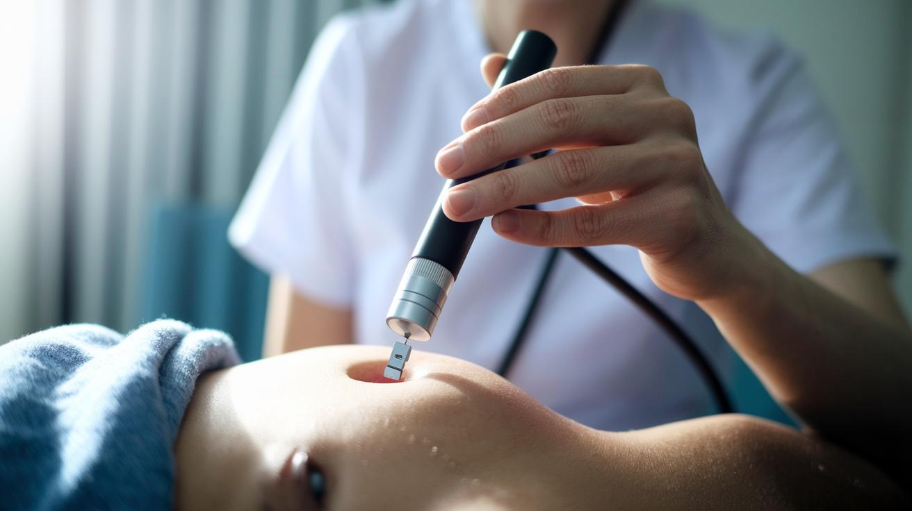
This test uses a thin tube (bronchoscope) to reach deep into your lung tissue. A small tool collects a tiny sample from areas that look unusual on live imaging. The tissue is then studied under a microscope to spot groups of immune cells called granulomas. Safety is a top priority during the test to avoid problems like bleeding. Doctors focus on finding noncaseating lesions (clusters of immune cells that do not have a dead center) to help tell sarcoidosis apart from infections or other conditions.
Extrapulmonary Biopsies (Skin/Lymph Node)
Sometimes, sarcoidosis affects other parts of the body outside the lungs. In these cases, doctors perform biopsies on the skin or lymph nodes. An ultrasound can guide the process to make sure the sample comes from the correct spot. The tissue is then handled carefully and sent for a detailed lab review. Special stains are used to rule out infections and other causes of granulomas. The lab report will clearly note if noncaseating granulomas are found. This detailed check helps confirm a diagnosis of sarcoidosis.
Clinician-reviewed • Last reviewed: October 2023
Differential Diagnosis and Exclusion in Sarcoidosis Confirmation
Before confirming sarcoidosis, your doctor needs to rule out other conditions that can look similar. They use a series of tests to make sure the diagnosis is correct and to avoid mistakes.
Your doctor often orders tests like cultures, blood tests (serology), imaging scans, and special tissue stains. These tests help pinpoint if something else is causing your symptoms.
They check for conditions such as:
- Tuberculosis
- Fungal infections
- Lymphoma
- Berylliosis
- Hypersensitivity pneumonitis
- Granulomatosis with polyangiitis
This step-by-step approach reviews both infections and autoimmune causes. By ruling out these possibilities, your doctor can be more confident that sarcoidosis is the cause of your signs and imaging findings. This careful process helps ensure you receive the right treatment.
Multispecialty Evaluation and Organ-Specific Confirmation in Sarcoidosis
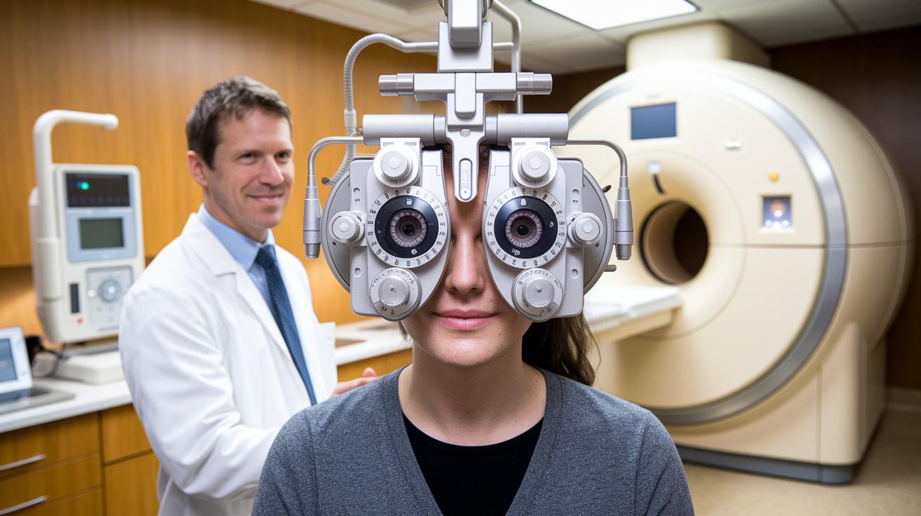
If you suspect sarcoidosis affects more than your lungs, your doctor may recommend an eye exam. An ophthalmologist checks for signs like pain, redness, or light sensitivity. This exam focuses on the front of your eye to spot inflammation (anterior uveitis). Finding these signs quickly and safely helps confirm that sarcoidosis may be affecting your eyes, which is key for planning the right treatment.
Heart tests are also important. Doctors use magnetic resonance imaging (MRI) and an electrocardiogram (ECG) to look for granulomas (small clusters of cells) and any issues with your heart’s electrical signals. The MRI can show scarring in the heart muscle, while the ECG catches any irregular heartbeats. These tests help uncover hidden heart problems and guide further treatment decisions.
A coordinated team of specialists then checks other areas of your body. A dermatologist, for example, may perform a skin biopsy if you have unusual bumps or plaques. You might also have neurological exams or assessments for lung and liver complaints. This team approach makes sure every possible area is reviewed so your treatment plan fits your needs.
Clinician-reviewed | Last reviewed October 2023
Final Words
In the action, this post outlined a clear roadmap for the diagnosis of sarcoidosis (how it is confirmed). It broke down the process, from assessing your history and symptoms to targeted imaging and lab tests. It explained the value of biopsies and provided ways to rule out other conditions. Each step builds your confidence as you track your symptoms and prepare for care. Keep these clear steps in mind as you move forward with a sense of clarity and control.
FAQ
Sarcoidosis diagnosis blood test
The sarcoidosis diagnosis blood test measures markers like ACE, calcium, and lysozyme. These levels, when combined with imaging and biopsy findings, help support a diagnosis.
How is sarcoidosis diagnosed?
The diagnosis of sarcoidosis involves a thorough history, physical exam, imaging tests, specific blood tests, and confirmatory biopsies that reveal noncaseating granulomas.
What are the sarcoidosis diagnostic criteria?
The sarcoidosis diagnostic criteria consist of clinical features, radiographic findings, lab tests, and a biopsy demonstrating noncaseating granulomas while ruling out other causes.
Is sarcoidosis cancer?
Sarcoidosis is an inflammatory disease, not cancer. It causes small clusters of cells called granulomas to form in various organs but does not involve cancerous growth.
How I cured my sarcoidosis?
Personal experiences of improvement vary. There is no single cure for sarcoidosis; treatment focuses on reducing inflammation and managing symptoms based on individual needs.
What are the current sarcoidosis diagnosis criteria?
The current criteria include a detailed history, physical exam, imaging studies, lab tests, and biopsy confirmation of noncaseating granulomas, along with exclusion of other similar conditions.
How to treat sarcoidosis?
Treating sarcoidosis usually involves careful monitoring and medications such as corticosteroids to reduce inflammation. The treatment plan is individualized based on disease severity and organ involvement.
Is sarcoidosis deadly?
Sarcoidosis is rarely deadly. While it can lead to complications in organs like the heart, most cases are managed successfully with proper medical care.
What diagnostic test confirms sarcoidosis?
A biopsy that shows noncaseating granulomas, combined with supportive clinical history, imaging, and lab tests, confirms the diagnosis of sarcoidosis.
What are the first signs of sarcoidosis?
The first signs of sarcoidosis often include fatigue, a dry cough, and shortness of breath. Some patients may notice skin rashes or eye irritation early on.
What could be mistaken for sarcoidosis?
Conditions such as tuberculosis, fungal infections, lymphoma, berylliosis, hypersensitivity pneumonitis, and granulomatosis with polyangiitis can be mistaken for sarcoidosis due to similar symptoms.
Does sarcoidosis show up on MRI?
Sarcoidosis can show up on MRI, especially in cases with cardiac involvement, where it may reveal areas of inflammation or scarring in the heart muscle.